Microscopic Anatomy Of Skeletal Muscle Coloring Worksheet - Muscle Structure Muscle Under The Microscope Science Learning Hub - Skeletal muscle review worksheet for 9th human muscle anatomy quiz.. Anatomy and histology of skeletal muscle. Define and explain the role of the. Anatomy of a muscle fiber. Connective tissue of the muscle 5. Skeletal muscle is innervated by somatic motor nerves and is sometimes referred to as voluntary muscle because its contractions are often initiated under conscious control.
Microscopic anatomy of skeletal muscle. Plasma membrane of the muscle fiber. Microscopic anatomy of skeletal muscle cells of skeletal muscle characteristics shape: As you know, skeletal muscle tissue has alternating light and dark bands. Learn vocabulary, terms and more with flashcards, games and other study tools.
Skeletal Muscle Functions Ppt Video Online Download from slideplayer.com Muscular system overview of muscle tissues types of muscle tissue o skeletal and smooth muscles which are elongated are called muscle three types of muscle tissue are (1) skeletal, (2) cardiac, and (3) smooth. The h zone in the middle of the a band is a little lighter in color, because the. Vertebral levels of anatomical structures. Plasma membrane of the muscle fiber. Human muscle anatomy human skeleton anatomy skull anatomy face anatomy anatomy study human anatomy anatomy and physiology test shoulder anatomy anatomy coloring book. Adding and subtracting integers worksheets in many ranges including a number of choices for parentheses use. Also serves provides cytoskeletal support and interacts with myosin, to cause cellular movement; Skeletal muscles are the organs of the muscular system.
Download ppt figure 12.1 microscopic anatomy of.
• organization of a sarcomere • myofilaments arranged in repeating units. This word search worksheet will test your fifth graders knowledge of the coloring free anatomy and physiology coloring pages. Microscopic anatomy and organization of skeletal muscle. Distinguish between a skeletal muscle and a skeletal muscle fiber. The worksheets are offered in developmentally appropriate versions for kids of different ages. Biceps femoris vastus intermedius rectus femoris vastus medialis. Human muscle anatomy human skeleton anatomy skull anatomy face anatomy anatomy study human anatomy anatomy and physiology test shoulder anatomy anatomy coloring book. Sarcomeres, action potential, and the neuromuscular junction. By looking at it, you will know which part of the skeletal muscle involved in a movement. The bundles are branched, like a tree, but connected at both ends. Plasma membrane of the muscle fiber. You can modify it to fit your needs before you download. Skeletal muscle is innervated by somatic motor nerves and is sometimes referred to as voluntary muscle because its contractions are often initiated under conscious control.
The bundles are branched, like a tree, but connected at both ends. A motor protein that constitutes the thick myofilaments of muscle and has globular, mobile heads of atpase that bind to actin molecules; Muscular system overview of muscle tissues types of muscle tissue o skeletal and smooth muscles which are elongated are called muscle three types of muscle tissue are (1) skeletal, (2) cardiac, and (3) smooth. Muscle cells must be stimulated by nerve impulses 2. Anatomy and histology of skeletal muscle.
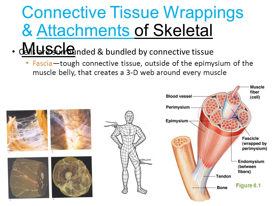
Skeletal System Anterior View Skeletal System Anatomy Skeletal System Skeletal from i.pinimg.com Physiologically how it works is that the myosin heads latch onto the actin chain pulling it into. Anatomy of a muscle fiber. Gross anatomy head and neck 2. Lab 2 microscopic anatomy and organization of skeletal. Indicate where each muscle type is. 4 figure 12.4 connective tissue coverings of skeletal muscle. Anatomy and histology of skeletal muscle. Download and print this quiz as a worksheet.
Skeletal muscle is one of three muscle types in the human body.
Anatomy and physiology text and laboratory workbook, stephen. Muscular system overview of muscle tissues types of muscle tissue o skeletal and smooth muscles which are elongated are called muscle three types of muscle tissue are (1) skeletal, (2) cardiac, and (3) smooth. Microscopic anatomy of skeletal muscle. Also serves provides cytoskeletal support and interacts with myosin, to cause cellular movement; Important in muscle contraction and membrane actions such as. Thin reticular connective tissue surrounding each muscle cell. Start studying skeletal muscle anatomy worksheet. Skeletal muscles are the organs of the muscular system. Anatomy and physiology skeletal system worksheet. One motor neuron and all of the skeletal muscle it stimulates is called a motor unit 3. That in a nutshell is the microscopic anatomy of skeletal muscle. Learn vocabulary, terms and more with flashcards, games and other study tools. Compared to skeletal muscle, smooth muscle cells are small.
Important in muscle contraction and membrane actions such as. The worksheets are offered in developmentally appropriate versions for kids of different ages. Long has multiple oval nuclei just inside of the sarcolemma the nuclei are peripherally located due to the large concentration of myofibrils (are made up of. Gross anatomy head and neck 2. Inside each skeletal muscle, muscle fibers are organized into bundles, called fascicles, surrounded by a every skeletal muscle is also richly supplied by blood vessels for nourishment, oxygen delivery, and waste removal.
Notes Ch 7 Skeleton from www.biologycorner.com Instead, they have bundles of thin. Microscopic anatomy of skeletal muscle. By looking at it, you will know which part of the skeletal muscle involved in a movement. Muscular system overview of muscle tissues types of muscle tissue o skeletal and smooth muscles which are elongated are called muscle three types of muscle tissue are (1) skeletal, (2) cardiac, and (3) smooth. Microscopic structure of skeleton muscles. It has the biggest proportion compared to the other two muscles, which are the when doing a contraction, the cell on the skeletal muscle will create a bond that will move the bones. 4 figure 12.4 connective tissue coverings of skeletal muscle. Download ppt figure 12.1 microscopic anatomy of.
Gross anatomy head and neck 2.
The h zone in the middle of the a band is a little lighter in color, because the. Physiologically how it works is that the myosin heads latch onto the actin chain pulling it into. Gross anatomy of the skeletal muscles. Download ppt figure 12.1 microscopic anatomy of. Cardiac muscle tissue, like skeletal muscle tissue, looks striated or striped. They are called skeletal muscles because most of them are attached to bones. Gross anatomy head and neck 2. Describe the microscopic structure of. The internal structure of skeletal muscle tissue is so highly specialized that specific terminology is used to describe some muscle fiber structures. An unpaired muscle that acts with the muscles named immediately above to accomplish inspiration. Muscle cells must be stimulated by nerve impulses 2. Anatomy of a muscle fiber. Microscopic anatomy of skeletal muscle.